Introduction
The demand for fisheries and aquaculture products has registered sustained growth since 1961, with an average annual increase of 3%. This global panorama is led by the dynamism of the aquaculture sector, whose production volume has progressively surpassed the yields of traditional capture fisheries. This industrial development and momentum directly respond to the increase in per capita consumption, which is determined by urbanization processes, rising purchasing power, and the evolution of food consumption trends on a global scale (FAO, 2022).
Within this scope, crustacean production occupies a significant fraction of total productive activity, with white shrimp (Litopenaeus vannamei) positioning itself as the fourth most produced aquaculture species globally and the second within the animal kingdom, surpassed only by the Pacific oyster (Crassostrea gigas) (FAO, 2022). White shrimp is a marine decapod crustacean native to the eastern coast of the Pacific Ocean, commonly associated with tropical waters. However, it tolerates a wide range of salinities, as well as moderate temperature variations and even low-salinity continental waters. This resilience, combined with its rapid growth and excellent feed conversion ratio, has driven its positioning as a key species for global aquatic production.
Despite its omnivorous habits, this species presents moderately high crude protein requirements, which usually range between 30% and 40% of the total feed composition, depending on the production stage and the stocking density of the production system (Jobling et al., 2012). Traditionally, the aquaculture sector has employed fishmeal as the main high-quality protein source to meet these needs. However, its market volatility and limited global supply have driven a strategic transition toward more sustainable and cost-effective plant protein sources, such as soy and corn derivatives, complemented with functional additives that optimize their digestibility.
Nevertheless, this modification in the formulation of aquaculture feeds has generated various challenges, among which a higher risk of exposure of fish to mycotoxins stands out as a priority.
Mycotoxins in aquaculture
Mycotoxins are toxic secondary metabolites generated by different species of fungi, which commonly contaminate both raw materials and finished feeds (Gruber-Dorninger et al., 2019). Their impact extends throughout the entire food chain, affecting humans directly through the ingestion of contaminated plant products, or indirectly through the consumption of animal-derived foods from livestock exposed to these toxins.
In the aquaculture sector, the exposure of cultured species is not restricted solely to the consumption of contaminated feed, since these compounds possess the capacity to persist in the water and sediments of production systems. This diversity in exposure pathways not only severely compromises animal health and production indices but also puts the food safety of the final consumer at risk due to the potential transfer of residues.
From the perspective of biological damage, the toxicity of these compounds triggers a complex clinical picture. The main described effects include cellular oxidative stress, severe histopathological lesions in target organs such as the gills and hepatopancreas, behavioral alterations, drastic drops in weight gain, and, in critical cases, high mortality. The severity of this symptomatology is variable and depends closely on the affected species, its developmental stage, the type of mycotoxins present, the ingested dose, and the exposure period (Oliveira et al., 2020).
Finally, a critical factor that aggravates this problem is the high thermostability of mycotoxins. This structural resistance allows them to effectively withstand the thermal treatments and extrusion processes common in aquaculture feed manufacturing plants, remaining active in the final feed (Gbashi et al., 2019; Sueck et al., 2019).
Mycotoxins in white Shrimp
The physiopathological effects of the most studied mycotoxins in white shrimp are detailed below. However, it is worth noting that beyond the individual action of these compounds, the most frequent scenario in nature is co-contamination by multiple mycotoxins rather than their isolated detection. The relevance of this phenomenon lies in the fact that these toxins usually exhibit synergism, meaning that the simultaneous presence of several in the same feed enhances their toxicity.
Aflatoxin B1
Aflatoxins are a group of mycotoxins produced by fungi of the genus Aspergillus, mainly A. flavus and A. parasiticus, and include variants B1, B2, G1, and G2. Among them, aflatoxin B1 (AFB1) stands out for its high carcinogenic potential and its tendency to bioaccumulate in target organs, such as the hepatopancreas and muscle tissue.
In white shrimp farming, exposure to AFB1 triggers a wide range of physiopathological effects that severely compromise production parameters. Research such as that by Yu et al. (2018) demonstrated that inclusions from 500 ppb in the diet induce a decrease in growth of up to 39.44% and an 11.25% drop in survival compared to animals fed mycotoxin-free diets; these effects become more acute with higher doses. This negative impact on performance is associated with digestive dysfunctions, alterations in the microbiota, oxidative stress, histological damage in the hepatopancreas, and immunosuppression.
Regarding gastrointestinal dysfunction, Su et al. (2021) reported a drastic decrease in the activity of key digestive enzymes (protease, lipase, and amylase) starting at doses of 2500 ppb. Similarly, under stress conditions caused by concentrations of 5000 ppb, Wang et al. (2018) evidenced a collapse of the intestinal microbiota. This imbalance was characterized by a decrease in beneficial bacterial groups such as Flavobacterium and Bacteroidetes, and a collateral increase in potential pathogens such as Vibrio and Photobacterium.
In relation to oxidative stress, the same study by Wang et al. (2018) confirmed an initial increase in the activity of antioxidant defense enzymes, such as superoxide dismutase (SOD), catalase (CAT), and glutathione peroxidase (GPX), starting at 5000 ppb, followed by a total collapse of the defensive system after two weeks of exposure. This drop was accompanied by an increase in malondialdehyde (MDA), a key biomarker of lipid peroxidation and cellular damage.
This pathological scenario is closely linked to structural alterations in the hepatopancreas. Su et al. (2025) reported the rupture of epithelial cells and an increase in the lumen starting from contaminations of 2500 ppb in the feed. Likewise, this hepatic damage is reflected in the plasma through an increase in biomarkers of cellular rupture, such as alanine aminotransferase (ALT) and aspartate aminotransferase (AST), according to observations by Yu et al. (2018).
Finally, the immunosuppression caused by this mycotoxin increases susceptibility to pathogenic agents. This immunological compromise manifests through an exacerbated inflammatory response, reflected in the plasma increase of inducible nitric oxide synthase (iNOS), and a notable reduction in the expression of genes essential for immunomodulation, such as those belonging to the Toll and Dorsal pathways.
Fumonisins
Fumonisins are mycotoxins originated by fungi of the genus Fusarium, mainly by the species F. verticillioides and F. proliferatum, which are responsible for the synthesis of variants B1, B2, and B3 (Cáceres et al., 2020).
In white shrimp cultivation, similarly to what is observed with AFB1, exposure to fumonisin B1 (FB1) severely compromises zootechnical parameters. Research such as that by Kracizy et al. (2021) demonstrated that dietary inclusions from 1715 ppb induce a 19.91% reduction in weight gain compared to animals fed contaminant-free diets.
In addition to affecting growth, this toxin alters the final quality of the product by destabilizing muscle proteins; García-Morales et al. (2015) reported reductions in the soluble protein of myofibrillar muscle and a lower water-holding capacity starting at doses of 200 ppb.
This negative impact becomes more acute upon reaching concentrations of 1000 ppb, presenting severe alterations such as the hydrolysis of myosin chains (Mexía-Salazar et al., 2008), which significantly reduces the shelf life of the product during storage.
Regarding tissue damage, various histopathological deteriorations have been recorded in target organs such as the hepatopancreas and gills. Mexía-Salazar et al. (2008) evidenced that contaminations of 500 ppb trigger the deformation and vacuolization of hepatopancreatic tubules, as well as initial signs of melanization. As exposure increases to doses of 1000 ppb, the damage progresses toward a marked infiltration of hemocytes and tissue necrosis. At this same inclusion level (1000 ppb), the authors found that the pathological impact extends to the gills, causing atrophy and severe anatomical damage.
Lastly, the compromise of the immune system manifests drastically starting from inclusions of 500 ppb. According to Mexía-Salazar et al. (2008), this concentration induces a severe reduction in the total circulating hemocyte count and decreases the production rate of the superoxide anion. This depresses phagocytic activity, a key mechanism in defense against pathogens. Likewise, a suppression of humoral immunity is observed at this same dose threshold, reflected in the drop of key immunological enzyme activities, such as phenoloxidase (PO).
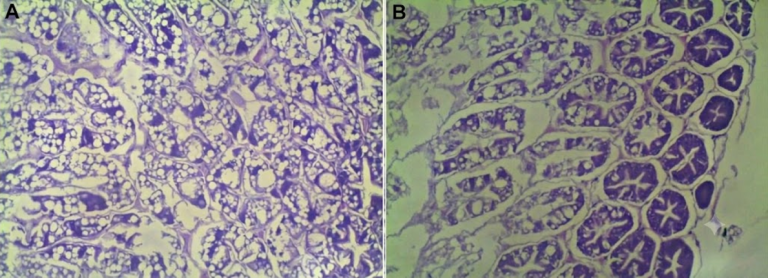
Figure 1. Histological sections of white shrimp hepatopancreas. (A) Intact tissue; (B) Cell atrophy and vacuolization by FB1 action (Kracizy et al., 2021).
Ochratoxin A
Ochratoxins constitute a group of fungal metabolites generated mainly by species of the genera Aspergillus and Penicillium. Within this group, ochratoxin A (OTA) stands out as the most prevalent variant and the one for which most effects have been recorded in aquaculture production species, including shrimp.
According to research by Albuquerque et al. (2016), exposure to OTA doses starting from 500 ppb is sufficient to induce significant drops in zootechnical performance. This negative impact shows a direct correlation with the increase of mycotoxin concentration in the feed: at a dose of 500 ppb, a 6% reduction in average weight gain is observed compared to healthy animals, as well as a deterioration in the feed conversion ratio, which increases from 1.01 to 1.09. At a dose of 1000 ppb, the conversion ratio worsens drastically until reaching a value of 1.23, while the growth rate experiences a contraction of 17.82% relative to the control group.
Parallelly, the study confirmed a worrisome phenomenon of carry-over of the toxin into tissues intended for human consumption. Specifically, the inclusion of just 100 ppb of OTA in the feed resulted in the retention of residues of 0.20 ppb in the abdominal musculature of the animals. This tissue accumulation not only seriously compromises the economic and productive viability of the batch but also introduces a direct risk to food safety and the health of the final consumer.
T-2 Toxin
T-2 toxin is a trichothecene produced by fungi of the genus Fusarium. These pathogens colonize corn, wheat, and soy crops, resulting in the contamination of grains that are subsequently used in the formulation of aquaculture feeds (Schatzmayr and Streit, 2013).
According to the findings of Qiu et al. (2016), exposure to a low dose of just 500 ppb is sufficient to drastically compromise productivity on farms. When evaluating diets contaminated with 500 ppb of T-2 toxin against mycotoxin-free controls, the following effects were observed: a decrease in the survival rate from 92.22% down to 78.88%; and weight gain slowed down significantly, descending from 22% to 18.30%.
This decline in productive performance is closely linked to a severe immunosuppressive effect, characterized by a drop in phenoloxidase (PO) activity, whose absorbance decreases from 17.93 min-1 × mg protein-1 in healthy animals to 12.09 and 9.61 with doses of 500 and 1200 ppb of the compound, respectively. Furthermore, a drastic reduction in the total hemocyte count is generated, which drops from 8.00 × 10⁶ to 5.00 × 10⁶ upon reaching 2400 ppb, leaving the organism unprotected against secondary pathogens.
At a physiological level, the hepatopancreas acts as the main target organ of toxicity, manifesting alterations that evidence the metabolic collapse of the digestive system.
The enzymatic evaluation performed by Qiu et al. (2016) demonstrates a plunge in the activity of glutamic-pyruvic transaminase (GPT), which is reduced from 62.73 U/L in the control diet to 25.03 U/L with a dose of 1200 ppb of T-2 toxin. Consistent with this biomarker of tissue damage, the integrated histopathological analyses of Qiu et al. (2016) and Bi et al. (2019) confirm a dose-dependent progression of lesions in the hepatopancreatic microstructure: at 500 ppb, they present with inflammatory processes, cellular atrophy, and a macroscopic reddish coloration. From 1200 ppb and 1500 ppb, the condition evolves toward marked cellular vacuolization; and at higher concentrations, it culminates in generalized cell lysis and severe necrosis of the tubular epithelium.
Simultaneously, the qualitative integrity of the muscle tissue and the lipid profile of the white shrimp suffer a systemic deterioration that depreciates the value of the final product. In a 20-day experimental design with 4 g specimens, Bi et al. (2019) determined that the ingestion of T-2 toxin alters the metabolism of body lipids, causing an initial reduction in the total content of saturated fatty acids (SFA), monounsaturated fatty acids (MUFA), and polyunsaturated fatty acids (PUFA) in the muscle under low-dosage conditions.
This chemical malnutrition correlates directly with damage to the crustacean’s meat, where microscopy reveals greater myofibrillar separation at doses up to 1500 ppb; a pathological state that aggravates exponentially from 4500 ppb up to 13500 ppb through the loss of tissue continuity, the separation of sarcomeres, and the loss of the compact structure of the muscle, negatively affecting the consistency and quality of the harvested biomass.
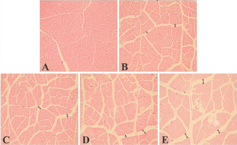
Figure 2. Histological slices of shrimp muscle with increasing doses of T2 toxin: (A) 0 ppb; (B) 500 ppb; (C) 1500 ppb; (D) 4500 ppb; (E) 13500 ppb. Increasing myofibrillar and sarcomere separation (Bi et al., 2019).
Deoxynivalenol
Deoxynivalenol (DON) is another highly relevant trichothecene also generated by fungi of the genus Fusarium. By colonizing agricultural crops such as corn and wheat, it contaminates the raw materials present in the feeds used in shrimp farming.
According to research conducted by Xie et al. (2018), the ingestion of deoxynivalenol induces a progressive penalty on the zootechnical indices of the crustacean. The results demonstrate a slight reduction in weight gain starting from a low inclusion of 250 ppb, an effect that aggravates significantly upon reaching higher concentrations. This decline in productive performance reflects a severe metabolic dysfunction and the energy cost assumed by the animal to try to counteract toxicity in its organism.
At a tissue level, deoxynivalenol exerts a dose-dependent action that destabilizes the digestive and functional barriers of the shrimp, with damage localized mainly in the hepatopancreas and intestinal tract. Histological evaluations by Xie et al. (2018) reveal that initial doses of 250 ppb cause hypertrophy in the B-cells of the hepatopancreas, a defensive cellular response that evolves negatively as the toxin increases to 500 ppb, where the fusion of intestinal mucosal folds, an increase in the diameter of hepatopancreatic tubules, and the appearance of cellular apoptosis are evidenced. Upon reaching the critical concentration of 1000 ppb, microscopic pathology reveals a scenario of advanced apoptosis, characterized by the massive presence of apoptotic bodies and notable swelling of the endoplasmic reticulum, confirming the collapse of the cellular machinery responsible for protein synthesis.
According to what was expounded by Xi et al. (2008), tissue damages are directly linked to oxidative stress and immunosuppression. The physiological response to the toxin depends strictly on the exposure level. From 500 ppb, cellular detoxification mechanisms are activated through the increase of the enzyme glutathione S-transferase (GST) and a higher expression of superoxide dismutase (SOD) and glutathione peroxidase (GPX).
However, the immune system shows a differentiated behavior according to the dose. While a low exposure of 250 ppb acts as an acute stressor that overstimulates the NF-kB pathway and the HSP70 protein to repair cellular damage, high doses of 1000 ppb cause the opposite effect. Face to a sustained challenge, this defense mechanism collapses, deriving in systemic immunosuppression and the over-activation of the prophenoloxidase (proPO) system.
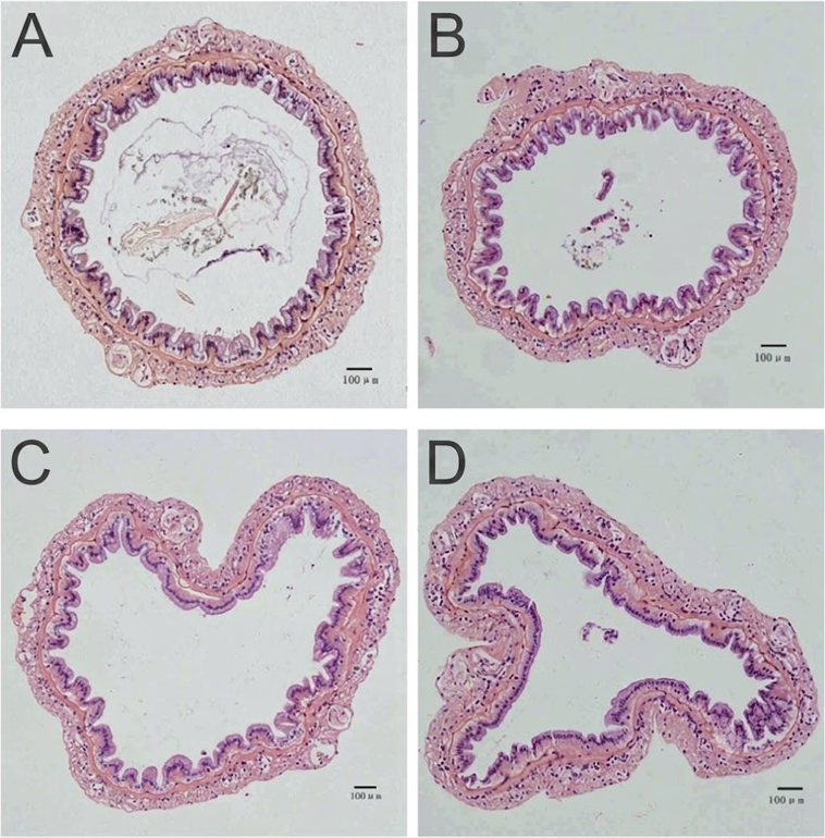
Figure 3. Histological sections of the intestine: (A) Intact tissue; (B, C, D) Structural and mucose alterations caused by increasing doses of DON (Xie et al., 2018).
Mycotoxins in other crustaceans
Although white shrimp usually concentrates most of the attention in aquaculture research due to its production volume, sensitivity to mycotoxins is not exclusive to this species. The impact of these fungal metabolites extends to other crustaceans of high commercial value, both marine and freshwater, manifesting through metabolic alterations, immunosuppression, and oxidative stress.
Tiger Shrimp
The tiger shrimp (Penaeus monodon) is a global aquaculture pillar thanks to its large size, rapid growth, and high value. Although its production suffered a sharp decline due to health crises compared to white shrimp, today it experiences a resurgence thanks to the development of pathogen-free genetic lines. In this way, it remains a key economic engine and a massive source of foreign exchange in tropical regions.
In the context of mycotoxins, Supamattaya et al. (2005) reported that prolonged exposure to OTA and DON during an eight-week period triggers specific physiological alterations in this species, differentially affecting its defensive and metabolic performance according to the administered dose.
When evaluating the effects of OTA at concentrations of 100, 200, and 1000 ppb, the most critical impacts manifested with the highest dose (1000 ppb), causing a significant decrease in the activity of the enzyme phenoloxidase (PO), which compromises the crustacean’s immune system, and a slight reduction in the body weight gain of the animals.
Conversely, DON showed a markedly different behavior when tested at doses of 500, 1000, and 2000 ppb, as it did not alter phenoloxidase activity or total hemocyte count.
Despite these differences and the absence of histopathological lesions in key tissues such as the gills, nervous tissue, or hepatopancreas, both mycotoxins shared a negative systemic impact. Supamattaya et al. (2005) identified a generalized reduction in plasma levels of alkaline phosphatase (ALP), glutamic-oxaloacetic transaminase (GOT), and glutamic-pyruvic transaminase (GPT). The drop in these hepatic and immunophysiological enzymes constitutes a crucial indirect indicator that both OTA and DON are capable of altering and deteriorating the proper cellular functioning of the hepatopancreas.
Chinese Mitten Crab
The Chinese mitten crab (Eriocheir sinensis) is one of the most valuable freshwater species in East Asia, characterized by strong traditional demand and gastronomic luxury. Its cultivation is carried out massively in lakes and ponds in China through semi-natural artificial propagation systems.
In the scope of its rearing, the analysis of feed contamination by mycotoxins is a critical factor not only for productivity but also for food safety, given the potential risk represented by the accumulation of toxins in its edible meat for human consumers. Recent investigations led by Yang et al. (2023) and Qiu et al. (2016) have shed light on the severe physiological effects that both T-2 toxin and AFB1 cause in this species, demonstrating highly damaging chronic and acute toxicity dynamics.
When evaluating T-2 toxin administered chronically for 56 days in juvenile crabs (at doses of 600, 2500, and 5000 ppb of feed), a direct impact on production indices was demonstrated, significantly reducing growth and weight gain at all evaluated concentrations, in addition to increasing mortality at the highest dose (5000 ppb). At a physiological level, this substance triggered a severe picture of oxidative stress, characterized by the elevation of malondialdehyde (MDA) and the plunge of antioxidant defenses superoxide dismutase (SOD) and glutathione peroxidase (GPX), combined with deep immunosuppression manifested in the drop of hemocyte count and respiratory burst. Likewise, the induction of inflammatory processes and apoptosis was found from the lowest dose, accompanied by severe morphological damage in the hepatopancreas that destroyed the basal membrane of its tubules and altered the cellular detoxification system.
On the other hand, the evaluation of the acute impact of AFB1 through a single injection of 400 µL (60 mg/L) revealed an immediate and intense toxicity concentrated in the hepatopancreas at 30 and 60 minutes post-exposure. This damage activated the expression of the sorbitol dehydrogenase gene, a direct marker of cell injury in this tissue, causing a strong immune and oxidative stress alteration.
Red Swamp Crayfish
The red swamp crayfish (Procambarus clarkii), native to North America, is the most cultivated crayfish species in the world due to its notable resistance, rapid growth, and high fertility. Its production leads freshwater aquaculture in countries like China and the United States, integrating successfully into crop rotation systems such as rice.
In the framework of the safety and immunity of this species, a study developed by Wen et al. (2023) evaluated the effects of exposure to DON through the administration of a dose of 3000 ppb. The treatment generated a notable physiological response at a defensive level, recording a metabolic increase in serum antioxidant enzymes superoxide dismutase (SOD), catalase (CAT), and glutathione S-transferase (GST), as well as an increase in the activity of the immune enzyme alkaline phosphatase (AKP).
Conclusion
The strategic shift toward plant-based proteins in aquaculture feeds has increased the exposure of white shrimp to mycotoxins. As detailed, the presence of toxins such as AFB1, FB1, OTA, T2, and DON triggers a multi-organ collapse characterized by oxidative stress, immunosuppression, severe histological damage in the hepatopancreas, and loss of muscle quality, which directly reduces the productive performance of farms.
Therefore, the rigorous management of mycotoxins through the use of efficient biotechnological solutions is an indispensable pillar to protect animal health, optimize zootechnical parameters, and guarantee the highest food safety standards required for human consumption.