Definition, etymology and general principles
Mycotoxins were first recognised over 5,000 years ago in China, but their effects were not scientifically documented until 1962, following a catastrophic veterinary crisis near London (United Kingdom), in which some 100,000 turkeys died (Blount, 1961). Subsequent investigations linked the outbreak to peanut meal contaminated with secondary metabolites produced by Aspergillus flavus (aflatoxins) and raised the possibility that other mould metabolites could also be toxic. This led to a broader classification of mycotoxins, encompassing compounds produced by well-known fungi, such as the ergot alkaloids of rye, substances originally isolated as antibiotics, like patulin, and a series of newly discovered secondary metabolites identified in moulds suspected of harming human or animal health, including ochratoxin A (Bennett et al., 2003).
Defining mycotoxins in a few words can be challenging. Mycotoxins are natural, low- molecular-weight compounds (that is, small molecules) produced as secondary metabolites by filamentous fungi. These metabolites form a toxigenic and chemically heterogeneous group that can cause disease and even death in humans and animals (Pitt et al., 2017).
The classification and definition of mycotoxins are particularly challenging, given the complexity of their chemical structures, the diversity of their biosynthetic pathways and biological activities, and their production by a broad range of fungal species.
To date, over 400 mycotoxins have been identified; however, only around a dozen groups are routinely monitored because of the risks they present to human and animal health (Agriopoulou et al., 2020). Although the reasons for their production are not yet fully understood, it is widely believed that they may serve as part of a fungal defence mechanism against insects, as many mycotoxins and their metabolites possess insecticidal properties (Richard, 2007).
Grains and cereals are particularly vulnerable to contamination with mycotoxin-producing fungi. Such contamination may occur during harvest (Diener et al., 1987), storage, or transport (Smith et al., 1985). When animals consume contaminated feed, exposure to mycotoxins can lead to severe toxic effects. Poisoning caused by mycotoxins is referred to as mycotoxicosis. Diagnosis generally requires demonstrating a dose–response relationship between the mycotoxin and the observed clinical signs.
The clinical signs of mycotoxicosis vary according to the type of mycotoxin, the amount and duration of exposure, and the species, age, health status, and sex of the exposed individual.
Mycotoxins adversely affect animal performance, health, and welfare, driving substantial research efforts in this area. Studies have explored the biosynthetic pathways of mycotoxins, their species-specific effects in livestock, and the associated clinical manifestations.
Characterisation and discovery of common mycotoxins
The discovery of this group of toxins dates back to the 1960s, when veterinarian William Percy Blount (Figure 1) investigated the deaths of 100,000 turkeys in the United Kingdom. After exhaustive testing of bacterial toxins, chemicals, pesticides, and every feed ingredient used, Blount (1961) ultimately traced the cause to a single source: the birds’ diet.
Although the link between the intoxication and the diet was confirmed, the precise cause remained unclear.

Figure 1. William Percy Blount.

Figure 2. Aspergillus flavus.
At the same time, an alarming increase in cases of liver disease among chickens was observed in Kenya, which ultimately led to the identification of peanut meal contaminated with Aspergillus flavus (Figure 2).
This discovery was made by J. J. Elphick of the Commonwealth Mycological Institute (UK), who identified the fungus by the pronounced fluorescence of aflatoxins (AFs) (Sargeant et al., 1961). Initial studies employed paper chromatography, which was later replaced by thin-layer chromatography (Coomes et al., 1963; Broadbent et al., 1963).
A decisive breakthrough in identifying the origin of AFs was facilitated by the geographical proximity of the affected turkeys to the Port of London, which enabled tracing the feed sources. This led to the identification of potentially contaminated raw materials, most notably the S.S. Rossetti meal, which had arrived from Brazil on 7 July 1960. In subsequent years, significant advances were made, including the development of more reliable analytical methods that allowed the identification of four AFs (B1, B2, G1 and G2) based on their fluorescent colours in thin-layer chromatography (Nesbitt et al., 1962; Pitt et al., 2017).
In 1920, mould-contaminated maize was occasionally associated with oestrogenic symptoms in pigs in the United States (McNutt et al., 1928). The same feed was administered to both guinea pigs and rats, resulting in uterine swelling in both species.
Christensen et al. (1965) succeeded in isolating the compounds present in the mouldy maize, identifying two distinct chemical structures, which they designated F-1 and F-2.
F-1 was identified as ergosterol and its identity was confirmed through various chemical reactions.
Urry et al. (1966) subsequently determined the chemical structure of F-2, later named zearalenone (ZEN) due to its structure and the fungus that produces it (Fusarium graminearum; Figure 3).
A synthetic form of the metabolite α-ZAL, known as zeranol, was used as an anabolic agent in sheep, sows, and other species in the United States (Diekman et al., 1989). However, in 1989, the European Union (EU) prohibited the use of zeranol.
Sows are highly sensitive to this mycotoxin, particularly because of the severe clinical signs affecting the reproductive system, as reported in the literature.
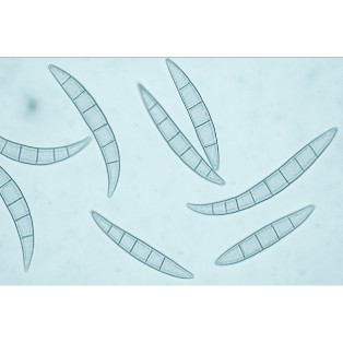
Figure 3. Fusarium graminearum.

Figure 4. Aspergillus ochraceus.
OTA was isolated and characterised in 1965 in South Africa as a metabolite of the fungus Aspergillus ochraceus (Figure 4), during a large-scale screening of fungal metabolites specifically aimed at identifying new mycotoxins (Van der Merwe et al., 1965).
This mycotoxin primarily affects the kidneys, with effects reported in all livestock species (Figure 5).
In addition, hepatic and immune alterations have also been reported, with OTA exhibiting potent immunosuppressive, teratogenic, and carcinogenic effects (Bennett et al., 2003).


Figure 6. Maize contaminated with Fusarium graminearum.
DON was discovered in the 1970s in Japan (Yoshizawa et al., 1973) as a metabolite of Fusarium graminearum (Figure 6). Subsequently, Vesonder et al. (1973) reported findings from a study conducted in pigs. Owing to outbreaks of emetic syndromes (clinical vomiting syndrome) associated with DON-contaminated feed, this mycotoxin is also referred to as “vomitoxin“.
When DON is ingested at high doses by farm animals, it causes nausea, vomiting and diarrhea, and at lower doses, pigs and other livestock show weight loss and feed refusal (Rotter et al., 1996).
Canadian researchers elucidated the toxigenic potential of Fusarium graminearum and identified two important chemotypes producing DON via either 15-acetyldeoxynivalenol or 3-acetyldeoxynivalenol (Greenhalgh et al., 1984; Kasitu et al., 1992).
The T-2 toxin, produced by Fusarium sporotrichioides (Figure 7), was first isolated in 1930 in the former Soviet Union and is known to induce the disease Alimentary Toxic Aleukia (ATA).
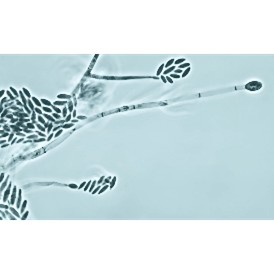
Figure 7. Fusarium sporotrichioides.
During the Second World War, labour shortages meant that forage crops were often left in the fields, leading to greater exposure to food made from contaminated wheat. Subsequently, Russian scientists identified the metabolites poaefusarin from Fusarium poae and sporofusarin from Fusarium sporotrichioides (Joffe, 1971). In addition, researchers at the University of Wisconsin (United States) isolated the T-2 toxin in 1966, which was later associated with the development of ATA (Mirocha et al., 1973).
Fumonisins were independently discovered by two research groups in 1988. One group was investigating the cause of oesophageal cancer in humans in certain regions of South Africa (Bezuidenhout et al., 1988), while the other sought to determine the aetiology of a well-known disease affecting horses, equine leukoencephalomalacia (ELEM) (Wilson et al., 1990). This family of mycotoxins is produced by species of the genus Fusarium, the best-known being fumonisin B1 (FB1) and fumonisin B2 (FB2), which can affect animals by disrupting sphingolipid metabolism (Figure 8).

Figure 8. Structure of sphingolipids.
High concentrations of FB1 and FB2 can induce porcine pulmonary oedema (PPE) (Colvin et al., 1992; Prelusky et al., 1994). At low concentrations, these toxins have been reported to cause liver and kidney damage in piglets (Riley et al., 1993). In chickens, fumonisins have been shown to induce intestinal damage (Javed et al., 1993).

Figure 9. Toxicity of fusaproliferin.
One of the first papers to use the term “emerging mycotoxins” was published in 2008, focusing on metabolites of Fusarium fusaproliferin (Jestoi, 2008). In a more recent publication, emerging mycotoxins were defined as “mycotoxins that are not routinely monitored, nor are they legislatively regulated; however, evidence of their occurrence is rapidly increasing” (Vaclavikova et al., 2013). This group comprises fusaproliferin, beauvericin, enniatins and moniliformin, all produced by Fusarium species, among other toxins.
Fusaproliferin (Figure 9), produced by Fusarium subglutinans and Fusarium verticillioides, was first isolated in 1995 by Ritieni. Available toxicity data are limited to Artemia salina larvae and chicken embryos, and include teratogenic effects (Logrieco et al., 1996; Ritieni et al., 1997).
Beauvericin (BEA; Figure 10) was first isolated in 1991 in the United States (Gupta et al., 1991). It has been reported to possess insecticidal, antimicrobial and antibiotic properties.
Although in vitro studies have shown that BEA is toxic to rodents and poultry, these effects have not been confirmed in vivo (Jestoi, 2008; Dornetshuber et al., 2009).
Enniatins were first described by Gaumann and his research group in 1947 from the fungus Fusarium orthoceras.
Subsequent studies identified a mixture of enniatins A (Figure 11), A1, B and B1 (Shemyakin et al., 1969), and a complex rich in enniatin A and A1 was shown to be the active component in an insecticidal fraction targeting spruce worm larvae (Strongman et al., 1988). Enniatins exhibit antibacterial and cytotoxic properties and can induce oxidative stress, which may in turn lead to secondary effects on animal health and productivity (Dornetshuber et al., 2009).

Figure 10. Structure of beauvericin.

Figure 11. Structure of enniatin.
This group of mycotoxins includes derivatives that are not detectable by conventional analytical techniques, as their chemical or molecular structure has been altered.
For example, in studies evaluating the accumulation of DON in maize, Miller et al. (1983) observed that the concentration of DON in field maize inoculated with Fusarium graminearum declined over the growing season. The authors speculated that this reduction could result from a chemical transformation of the toxin by the plant’s metabolism. Shortly thereafter, it was reported that the DON content of yeast-fermented food products exceeded that of the contaminated wheat flour used in their production (Young et al., 1984). This increase was attributed to the enzymatic conversion of a DON conjugate produced by the wheat plant into DON-3-glucuronide (DON-3-GlcA), deepoxy-DON (DOM-1) and DON-15-glucuronide (DON-15-GlcA) (Figure 12).

Figure 12. DON metabolism.
ZEN can be modified by microorganisms and plants; however, these modified compounds can be fully converted back to ZEN by the intestinal microbiota in animals or humans. ZEN metabolism can be divided into two phases: Phase I and Phase II metabolism (Yang et al., 2017). Zearalenone-14-sulfate (ZEN-14-S) was identified in crops affected by Fusarium graminearum in 1991 (Gareis et al., 1990) as a product of ZEN metabolism (Figure 13).

Figure 13. Structure of ZEN-14-S.
Conclusion
Although the harmful effects of mycotoxin exposure have been studied for many years, their metabolic mechanisms of action remain under investigation, as do the strategies needed to mitigate their impact on animal health and productivity, the profitability of agricultural operations, and food safety.