Absorption, laminar necrosis, and the silent link in gastric toxicosis
Impact of mycotoxins on the ruminant's gastric compartments
Introduction
The omasum is the gastric compartment of the ruminant that receives the least clinical and scientific attention compared to its neighbors: the rumen absorbs most of the interest of nutritionists, and the abomasum monopolizes the attention of clinicians due to high-incidence pathologies such as displacement or ulceration. The omasum, quietly hidden between the reticulum and the abomasum, performs functions of water absorption, digesta concentration, and pH buffering that, when compromised, produce consequences that are difficult to attribute directly to this compartment in everyday practice (Mattioli, 2003; Mostrom & Jacobsen, 2020).
This diagnostic invisibility is amplified when the cause of the damage is mycotoxins. Omasal lesions induced by trichothecenes, aflatoxins, and Aspergillus metabolites are histologically documentable at necropsy, but clinically silent in their early stages. The animal shows no omasum-specific signs: the loss of function is expressed as diffuse metabolic deterioration, osmotic diarrhea, reduced absorption of amino acids and electrolytes, and worsening of conditions originating in the rumen and reticulum (Gallo et al., 2015). In this sense, the omasum is the silent link in the chain of gastrointestinal damage caused by mycotoxins.
This article reviews the anatomy and histology of the omasum in the context of its vulnerability to mycotoxins, the mechanisms of epithelial damage induced by trichothecenes and Aspergillus metabolites, the consequences for water-electrolyte balance and the preparation of digesta for abomasal digestion, and the practical implications for diagnosis and prevention.
1. Anatomy and histology of the omasum
The omasum is a spherical or ovoid organ located to the right of the midline, cranioventrally, immediately lateral to the reticulum and the ventral rumen, as shown in Figure 1. In adult cattle it has a capacity of 10-20 liters; in sheep and goats it is proportionally much smaller (1-2 liters). In camelids (camels, llamas, alpacas) the omasum is practically absent or highly rudimentary, which constitutes a significant functional difference compared to true ruminants (Clauss & Hummel, 2017; Mattioli, 2003).
Internally it presents between 100 and 130 laminae (longitudinal folds) of variable size that project from the wall toward the lumen of the organ, creating a labyrinth of channels. The free surface of the laminae is densely covered with keratinized papillae, shorter and sturdier than those of the rumen. The total surface area of the omasum can reach several square meters in adult cattle. The mucosa is lined by parakeratinized stratified squamous epithelium, histologically identical to that of the rumen and reticulum. The muscular wall between the laminae is especially well developed, with layers that allow active compression of the content trapped between them (Mattioli, 2003; Annison & Bryden, 1998).
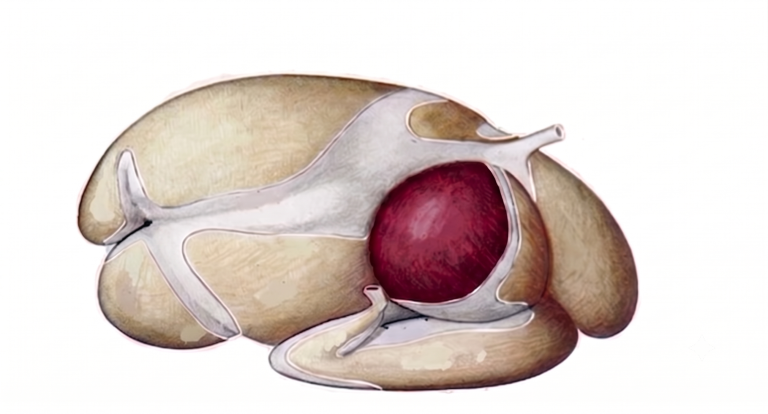
Figure 1. Lateral view of the forestomachs of a ruminant with emphasis on the omasum (colored). Adapted from Popesko (1977).
2. Physiology of the omasum
2.1. Absorption of water, electrolytes, and residual VFAs
The primary function of the omasum is the active and passive absorption of water, sodium, potassium, magnesium, and chloride, as well as of the VFAs that were not absorbed in the rumen. The content arriving from the reticulum has a semi-liquid consistency. As it advances between the omasal laminae, the mucosa actively extracts the water and electrolytes, producing progressive dehydration of the digesta. It is estimated that the omasum can absorb between 60% and 70% of the water entering from the reticulum, making it an organ of major importance for water conservation (Armstrong & Beever, 1969; Annison & Bryden, 1998).
This digesta-concentrating function serves a dual physiological purpose: it recovers large volumes of water and electrolytes, reducing the osmotic load on the abomasum and intestine; and it concentrates the protein material (bypass protein and microbial biomass) that must be enzymatically digested in the abomasum, optimizing the efficiency of subsequent acid hydrolysis and enzymatic hydrolysis.
2.2. Filtration and pH transition
The reticulo-omasal orifice, of small diameter (2-3 cm in cattle), acts as a mechanical filter that prevents the passage of large particles. The omasum also participates in buffering the pH of the digesta, smoothing the transition between the neutral-alkaline environment of the rumen (pH 5.5-7.0) and the strongly acidic environment of the abomasum (pH 2.0-3.5) (Mattioli, 2003; Yu et al., 2026).
3. Impact of mycotoxins on the omasum
3.1. Epithelial cytotoxicity: necrosis of the omasal laminae
The omasum, with its enormous absorptive surface formed by hundreds of laminae covered with keratinized papillae, represents a tissue especially susceptible to mycotoxin-induced epithelial damage. Trichothecenes, particularly T-2 toxin and DON, are potent inhibitors of ribosomal protein synthesis, triggering apoptosis and necrosis in the epithelium lining the omasal laminae. Added to this damage is the effect of aflatoxin B1 (AFB1), which drastically reduces the functional surface available for the absorption of water, sodium, potassium, and magnesium, compromising the water and electrolyte balance of the entire organism (Antonissen et al., 2014; Mostrom & Jacobsen, 2020).
3.2. The omasum as a target organ in bovine aspergillosis
In necropsies of cattle with Aspergillus mycotoxicosis, the omasum has been identified as the main target organ of the invasive fungal infection, being affected even before the reticulorumen and the abomasum. Gliotoxin, an immunosuppressive metabolite produced by Aspergillus fumigatus, induces apoptosis of lymphocytes and macrophages in the omasal mucosa, compromising the local immune response and allowing colonization of tissues by opportunistic fungi. This anatomical susceptibility has direct practical implications: histological damage to the omasum is not easily detectable in early stages by routine clinical diagnostic methods, so the functional loss tends to be subclinical and contributes to the silent deterioration of body condition and productivity (Gallo et al., 2015).
3.3. Consequences of absorptive failure
Because toxins such as fumonisins (FBs) and zearalenone (ZEN) show minimal ruminal degradation or become concentrated during dehydration of the digesta, they reach the abomasum while retaining their biological activity intact. This compromise of the omasum’s absorptive function has multiple nutritional consequences. The reduction in water absorption causes content to reach the abomasum with greater dilution, altering the concentration of the substrate for proteolytic enzymes and hydrochloric acid. The reduced absorption of sodium and potassium predisposes the animal to osmotic diarrhea and hyponatremia. The residual VFAs that were not absorbed in the rumen or the omasum reach the abomasum in abnormal proportions, altering the pH and the sequence of enzymatic activation (Annison & Bryden, 1998; Gao et al., 2020).
The disruption of omasal function represents the intermediate link in an axis of progressive damage that leads to suboptimal enzymatic digestion in the abomasum and to deficient intestinal absorption of amino acids and glucose. This deterioration, added to that already established in the rumen and reticulum, completes the picture of malabsorption and chronic energy deficit characteristic of subclinical mycotoxicosis in high-producing ruminants.
4. Practical relevance
Since omasal damage caused by mycotoxins is histological and subclinical in its early stages, in vivo diagnosis requires an indirect approach: monitoring of water and electrolyte balance (blood and urine analysis), assessment of fecal consistency (persistent mild osmotic diarrhea as a warning sign), systematic mycotoxin analysis of raw materials and total mixed rations (TMR), and systematic necropsy of animals that die or are culled from the herd to detect characteristic omasal lesions. Integrating these indicators makes it possible to identify situations of subclinical mycotoxicosis before the damage spreads and becomes established (Mostrom & Jacobsen, 2020).
Conclusion
The omasum is the most silent compartment of the ruminant gastric system, but its damage from mycotoxins has far-reaching nutritional and metabolic consequences. Epithelial necrosis of the omasal laminae induced by trichothecenes and metabolites of Aspergillus compromises the absorption of water and electrolytes, impairs the preparation of digesta for abomasal digestion, and amplifies the energy deficit initiated in the rumen. Recognizing the omasum as a specific target of mycotoxins is essential for building a complete model of the impact of these contaminants on ruminant digestive health.
To mitigate this damage, it is essential to implement bioprotective solutions for mycotoxin mitigation. These strategies help protect the integrity of the omasal laminae and preserve their absorptive capacity, ensuring the stability of the water balance and the overall digestive efficiency of the animal.