Introduction
Reproductive efficiency is one of the key economic determinants of cattle, sheep, and goat production: it determines the calving/lambing interval, the number of calves or lambs born per year, herd replacement rates, and, in dairy cattle, production persistency. However, subfertility and infertility are among the most frequent and economically costly health problems in ruminant production systems globally. Their clinical manifestations are diverse: repeat breeding, prolonged postpartum anestrus, ovarian cysts, subclinical endometritis, early embryonic mortality, abortions, and, in males, reduced semen quality and reduced libido.
The causes of these disorders are multifactorial and often coexist under field conditions: nutritional and energy imbalances, especially during the periparturient period; infectious diseases of the reproductive system (such as brucellosis, campylobacteriosis, trichomoniasis, or bacterial metritis); genetic and management factors; heat stress; and, with increasing scientific evidence, chronic exposure to mycotoxins present in the diet (Hartinger et al., 2022; Ogunade et al., 2018; Liu et al., 2023). For a long time, the latter factor was underestimated in ruminants, based on the assumption that ruminal fermentation acted as a sufficient protective barrier against these contaminants (Fink-Gremmels, 2008). Research over the past two decades has substantially challenged this assumption.
Mycotoxins are secondary metabolites produced by filamentous fungi, mainly by fungi of the Aspergillus, Fusarium, and Penicillium, which contaminate forages, silages, and their raw materials both in the field (preharvest contamination, favored by drought, excessive moisture, or insect damage) and during storage (inadequate drying, inadequate ventilation, residual moisture) (Dellait Animal Nutrition and Health, n.d.). It is estimated that a high proportion of grains and forages produced worldwide contain detectable concentrations of one or more mycotoxins, and the coexistence of multiple toxins in the same feed, known as multiple contamination or the “mycotoxin cocktail”, is a common occurrence under field conditions, which complicates diagnosis and assessment of the actual risk to the herd.
Although for decades it was assumed that ruminants were relatively protected owing to the ability of the ruminal microbiota to degrade these compounds, it has now been shown that this protection is partial: some mycotoxins resist ruminal metabolism, while others are converted into metabolites that are equally or more toxic than the original molecule. In addition, ruminal detoxification capacity may decrease under conditions associated with higher reproductive risk, such as high-concentrate diets, a low forage proportion, or high-producing animals with shorter ruminal retention times (EFSA, 2017; Fink-Gremmels, 2008).
The reproductive system is among the physiological systems most sensitive to this exposure, in both females and males: mycotoxins can act as endocrine disruptors, generate oxidative stress in gametes, impair follicular and luteal function, alter the uterine environment, and directly affect the early-stage embryo (Guerrero-Netro et al., 2015; Pizzo et al., 2016). The resulting condition is often subclinical and difficult to distinguish from other causes of infertility, and can easily be confused with other causes of infertility, and which, due to its insidious nature, is often detected only once it has already had a measurable impact on the herd’s reproductive indicators and, consequently, on farm profitability (Fushimi et al., 2015).
This article reviews the mechanisms by which the main mycotoxins affect reproduction in ruminants, synthesizes the available experimental and field evidence, and proposes diagnostic and control strategies applicable at the farm level.
The ruminal microbiota: an incomplete barrier
The rumen acts as an initial metabolic barrier against ingested mycotoxins. Ruminal protozoa and bacteria are capable of completely or partially degrading compounds such as zearalenone (ZEN) or deoxynivalenol (DON) (EFSA, 2017). However, this process does not necessarily constitute true detoxification: ruminal biotransformation of ZEN generates α-zearalenol, a metabolite with estrogenic activity several times higher than that of the original molecule, and β-zearalenol, which has direct toxic effects on the endometrium (EFSA, 2017; Fink-Gremmels, 2008). In other words, ruminal metabolism can sometimes enhance adverse reproductive effects rather than neutralize it.
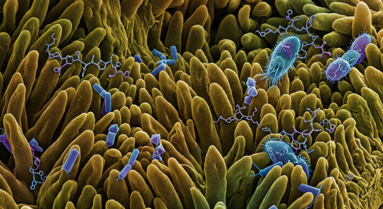
Figure 1. Interaction of the ruminal microbiota with mycotoxins ingested through the diet. Ciliated protozoa and various ruminal bacterial populations act together and may degrade, hydrolyze, or metabolize toxic compounds.
Main mycotoxins involved and their reproductive effects
Zearalenone (ZEN)
Zearalenone has the clearest and best-documented link to reproductive dysfunction in ruminants. Produced by Fusarium species, it has a chemical structure similar to that of endogenous estrogens, so it acts as an endocrine disruptor by binding to estrogen receptors (EFSA, 2017).
In females, exposure is associated with clinical signs such as (Fushimi et al., 2015; Pizzo et al., 2016; de Almeida Silva et al., 2021):
- Alterations in the estrous cycle (irregular or prolonged estrus and anovulatory cycles).
- Formation of estrogenically active cystic follicles.
- Vulvar hypertrophy and inflammation and, in severe cases, vaginal or uterine prolapse in non-pregnant animals.
- Reduced corpus luteum size and alterations in the antral follicle population.
- Lower oocyte quality in heifers.
- Increased embryonic mortality and abortion risk.
In young males, signs of feminization, mammary hypertrophy, and reduced circulating testosterone concentrations have been described. In adult bulls, however, cases of infertility associated with toxin-induced oxidative stress have been described (Liu et al., 2023).
Field observations suggest that ZEN concentrations above 250 µg/kg in the total ration may be associated with reproductive disorders, while levels close to 500 µg/kg have been associated with clinically evident hyperestrogenism. However, the response varies depending on the physiological status of the animal: some studies in postpartum cows have found no clear effects on fertility despite measurable follicular alterations (Fushimi et al., 2015), indicating that dose, duration of exposure, and production stage modulate the severity of the condition.
Fushimi et al. (2015) evaluated postpartum dairy cows exposed to ZEN concentrations below the permitted contamination threshold in Japan and found measurable alterations in the ovarian antral follicle population, although without a statistically significant effect on overall fertility in the group, suggesting that subclinical damage can precede the manifestation of overt infertility.
A controlled trial in non-lactating cows fed a diet containing approximately 25 ppm of ZEN showed a significant reduction in corpus luteum size compared with the control group, supporting a direct effect on luteal function.
Almeida Silva et al. (2021), cited in a study published in the Journal of Dairy Science (2025), reported that even relatively low ZEN concentrations (0.3 mg/kg of diet) were associated with reduced oocyte quality in cattle, although without significantly affecting the subsequent rate of embryonic development, suggesting a differential threshold between damage to the oocyte and damage to the developing embryo.
In contrast, Takagi et al. (2008) found no effect of elevated ZEN concentrations on in vitro fertilization or embryonic development rates, underscoring the variability of results depending on experimental design, route of exposure, and the stage of the reproductive cycle evaluated.
In vitro studies on granulosa cells from small bovine follicles (Pizzo et al., 2016) showed that ZEN and its metabolites, either alone or in combination with DON, alter cell proliferation, steroid production, and the expression of genes related to steroidogenesis.
Under field conditions, a 2024 study published in Mycotoxin Research that monitored urinary biomarkers of ZEN and beta-zearalenol in dairy herds reported that urinary ZEN concentrations above 2.7 ng/mL were associated with a significantly longer interval until the next pregnancy, a finding originally reported in Canadian farms (Tazerout, 2016) and subsequently evaluated in a new sampling campaign.
Deoxynivalenol (DON) and trichothecenes
DON and other trichothecenes (T-2 toxin, HT-2, diacetoxyscirpenol, nivalenol), also of Fusarium origin, mainly affect gastrointestinal and immune functions, but their reproductive impact is largely indirect: immunosuppression, reduced feed intake, and deterioration of overall metabolic status compromise reproductive efficiency by delaying the resumption of postpartum cyclicity and affecting the quality of the uterine environment (Ogunade et al., 2018; Guerrero-Netro et al., 2015). At the follicular level, DON also has a direct effect on the steroidogenesis of granulosa cells.
Guerrero-Netro et al. (2015), in a serum-free culture system with bovine granulosa cells, showed that DON inhibits estradiol and progesterone secretion, reduces the expression of the CYP19A1 gene (aromatase, a key enzyme in estrogen synthesis), and increases apoptosis through activation of the MAPK pathways (phosphorylation of p38 and JNK), providing a plausible molecular mechanism for the follicular dysfunction observed in the field.
A 2024 study on mycotoxin metabolites (deepoxy-deoxynivalenol and beta-zearalenol) evaluated their direct effects on the development of preimplantation bovine embryos, based on the premise that early embryonic mortality is the most common cause of reproductive failure in dairy cattle, with decreases in pregnancy rates of up to 20–30 percentage points between day 0 and day 7 of embryonic development (Gačnikar et al., 2024).
A two-year longitudinal study in European dairy herds, published in Journal of Dairy Science (2023), associated feeding management and combined exposure to Fusarium mycotoxins with alterations in udder health, production performance, and herd fertility, reinforcing that under practical farm conditions the effects are rarely due to a single mycotoxin in isolation.
A field study specifically designed to evaluate the combined effects of ZEN, DON, and fumonisin B1 on 486 Holstein cows analyzed blood metabolite concentrations, milk production, estrous activity, pregnancy rate, and uterine health. During the contamination period, a high proportion of cows with clinical metritis and a higher incidence of ovarian follicular cysts was recorded, and the authors note that co-exposure to several mycotoxins may play an important role in cystic ovarian disease, with herd recovery requiring a prolonged period after exposure has ceased.
Aflatoxins (AFs)
Produced by Aspergillus flavus and A. parasiticus, the main concern in dairy ruminants is their transfer to milk as aflatoxin M1 (AFM1), with implications for food safety (Hartinger et al., 2022). From a reproductive perspective, their hepatotoxicity indirectly compromises fertility by altering the metabolism of sex steroids and causing immunosuppression. The offspring of exposed animals may show greater susceptibility to inflammatory processes and secondary infections (Hartinger et al., 2022; Liu et al., 2023).
In water buffalo, dietary administration of AFs to water buffalo Bulls reduced the percentage of live spermatozoa from 91.1% to 80.8% and increased the proportion of abnormal spermatozoa to 54%, with semen evaluated one, four, and eight weeks after exposure (Hafez et al., 1982).
In Merino rams, a 92-day trial involving dietary AF exposure showed that, from the third week onward during liquid semen storage at 5 °C, the exposed group had significantly lower sperm motility and a higher proportion of abnormal and non-viable spermatozoa compared with the control group, an effect that was partially reversed by supplementation with esterified glucomannan as a mycotoxin-sequestering agent (Ataman et al., 2014).
In cattle, a study published in Toxicology (2017) exposed spermatozoa from bull ejaculates and epididymides to increasing concentrations of aflatoxin B1 (AFB1) (0.1 to 100 µM) and found reduced sperm viability, mitochondrial membrane hyperpolarization, and increased sperm DNA fragmentation even after short exposures, along with reduced fertilizing capacity during in vitro fertilization.
A follow-up study in the journal Reproduction (2020) analyzed the proteome of AFB1-treated bovine spermatozoa and the transcriptome of the resulting embryos, showing that fertilization with apoptotic spermatozoa exposed to the toxin resulted in no blastocyst formation, while fertilization with the non-apoptotic sperm subpopulation resulted in reduced cleavage rates and blastocysts with altered expression of key genes involved in early embryonic development (POU5F1 and SOX2), demonstrating a paternal gamete-to-embryo effect, as shown in Figure 2.
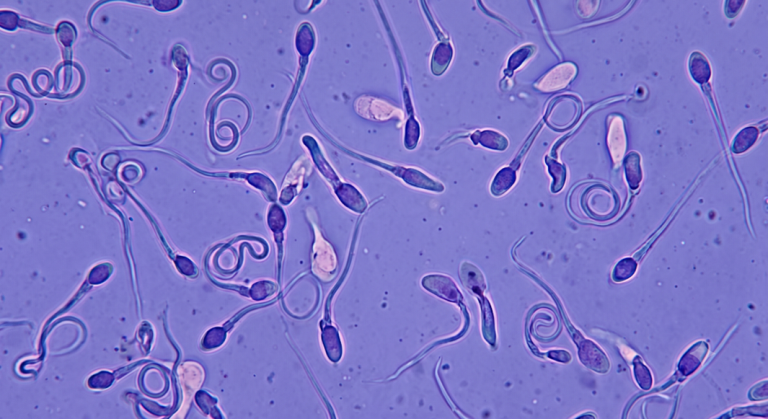
Figure 2. Microscopic alterations in male gametes. The alterations associated with AF and ZEN exposure are illustrated. A high proportion of spermatozoa exhibit with morphological abnormalities: deformed sperm heads, coiled or fragmented tails.
Fumonisins (FBs)
Fumonisins undergo slow and incomplete metabolism in the rumen, which may contribute to hepatotoxic and nephrotoxic effects (Fink-Gremmels, 2008). Their reproductive effects are less specific than those of ZEN, but they contribute to the deterioration of overall body condition and, therefore, to a decline in herd reproductive efficiency (Ogunade et al., 2018).
Ochratoxin A (OTA)
OTA is less studied in ruminants than in monogastrics because of its relatively efficient ruminal degradation, although under conditions of high or prolonged exposure it can contribute to nephrotoxicity and metabolic deterioration that indirectly affects reproductive function (Fink-Gremmels, 2008).
General pathophysiological mechanisms
The reproductive effects of mycotoxins in ruminants can be explained by several pathways, often acting simultaneously:
- Direct endocrine disruption: particularly through ZEN and its metabolites, which mimic the action of estrogens and alter the hypothalamic-pituitary-gonadal axis (EFSA, 2017; Pizzo et al., 2016).
- Oxidative stress: damage to oocytes, spermatozoa, and testicular tissues, thereby compromising gamete quality (Liu et al., 2023).
- Immunosuppression: increased susceptibility to uterine infections and mastitis, which can delay uterine involution and conception (Ogunade et al., 2018; Hartinger et al., 2022).
- Ruminal and hepatic dysfunction: reduced overall metabolic efficiency, potentially resulting in a negative energy balance, a recognized contributor to postpartum subfertility (Fink-Gremmels, 2008).
- Transgenerational effects: offspring with an increased predisposition to inflammatory processes (Hartinger et al., 2022).
These reproductive consequences are reflected in concrete reproductive performance indicators: a longer calving interval, a greater number of services per conception, a higher rate of repeat breeding, a greater incidence of ovarian cysts, and embryonic losses; as shown in Figure 3. In addition, mycotoxin contamination is usually accompanied by a decline in milk production and quality, thereby worsening the overall economic impact on the farm.
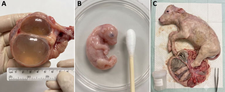
Figure 3. A) Macroscopic view of a bovine ovarian structure with large follicles with estrogenic activity. B) Bovine embryo recovered at an early developmental stage. C) Aborted bovine fetus together with its fetal membranes in a veterinary diagnostic laboratory setting.
Diagnosis and risk assessment
Identifying mycotoxin exposure as a cause of subfertility is complex, since its clinical signs overlap with those of other nutritional, infectious, and management-related conditions (Hartinger et al., 2022). A sound approach should combine: rapid screening tests for low-fiber raw materials, mold enumeration, and specific mycotoxin analyses (ideally by LC-MS/MS, which allows multiple mycotoxins to be quantified with high sensitivity) in fibrous ingredients and in the total mixed ration (TMR), together with correlation of the analytical findings with the herd’s reproductive and productive records.
Conclusion
Although ruminants have a limited capacity to degrade mycotoxins in the rumen, this protection is insufficient against compounds such as ZEN, whose estrogenic activity may even be enhanced by ruminal metabolism, thereby directly compromising reproductive function of both females and males. Addressing this problem requires a comprehensive strategy that combines agronomic preventive measures, regular analytical monitoring of the diet, and monitoring of the herd’s reproductive performance indicators, since diagnosis based exclusively on clinical signs is often late and nonspecific.