Glandular injury, ulcers, compromised proteolysis, and food safety
Impact of mycotoxins on the ruminant's gastric compartments
Introduction
The abomasum is the only gastric compartment of the ruminant that possesses true glandular tissue, and is histologically and functionally equivalent to the simple stomach of monogastrics. It is in the abomasum that the chemical and enzymatic preparation of the digesta is completed: the parietal cells secrete hydrochloric acid at a pH of 2.0-3.5, the chief cells release pepsinogen, which is converted into pepsin, and the G cells of the pyloric antrum produce gastrin to regulate the process. The integrity of this secretory system is an indispensable condition for the proper digestion of the microbial protein coming from the omasum, the main source of essential amino acids for the adult ruminant (Annison & Bryden, 1999; Mattioli, 2003).
When mycotoxins manage to overcome or evade the ruminal detoxification mechanisms, which occurs consistently with aflatoxin B1 (AFB1), deoxynivalenol (DON), T-2 toxin, and their metabolites, and reach the abomasum, they act on an epithelium already subjected to the most extreme physicochemical conditions of the entire digestive tract. The acidic pH, far from protecting the tissue, facilitates the activation of some toxins and the denaturation of the mucosa’s protein defenses. The result is glandular damage that adds to that already established in the rumen, the reticulum, and the omasum, representing the final station of a progressive and integrated process of degradation.
In addition to its local effects, the abomasum has a unique relevance in the context of food safety: the transfer of aflatoxin M1 (AFM1) to bovine milk, a consequence of the hepatic metabolism of the AFB1 absorbed from the digestive tract, is internationally regulated due to its implications for public health. This dimension means that the study of the impact of mycotoxins on the abomasum extends beyond the veterinary field to become a matter of food safety.
1. Anatomy and histology of the abomasum
The abomasum is located in the right ventral region of the abdominal cavity, from the level of the tenth rib to the abdominal floor. In adult cattle it has a capacity of 15-30 liters. Its shape is elongated and curved, with a cranial fundus and a caudal pylorus that connects it to the duodenum, as shown in Figure 1. In high-producing cows it can shift into abnormal positions (left or right displacement), a condition with high incidence around calving (Smith, 2023; Mattioli, 2003).
The abomasal mucosa is the only one of the four compartments that possesses glandular tissue. It is lined by a simple mucus-secreting columnar epithelium. In the lamina propria, three regions are distinguished: (1) cardiac glands, which produce mucus; (2) fundic glands, with parietal (oxyntic) cells producing hydrochloric acid (HCl) and intrinsic factor, chief (zymogenic) cells producing pepsinogen, and mucous neck cells; and (3) pyloric glands, rich in G cells producing gastrin (Mattioli, 2003; Annison & Bryden, 1999).
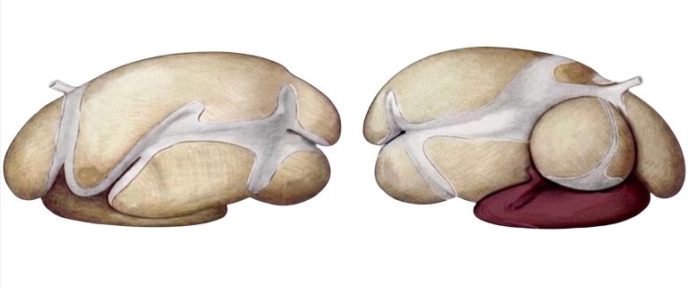
Figure 1. Lateral view of the forestomachs of a ruminant with emphasis on the abomasum (colored). Adapted from Popesko (1977).
2. Physiology of the abomasum
2.1. Acid digestion and protease activation
The parietal cells secrete HCl via a proton pump (H⁺/K⁺-ATPase), establishing an intraluminal pH of 2.0-3.5 in the adult animal. This acidity denatures proteins, making them susceptible to proteolytic hydrolysis, activates pepsinogen by converting it into active pepsin, and exerts a bactericidal effect that controls microbial growth in the posterior segments of the digestive tract. Gastrin, secreted by the G cells in response to distension and the presence of peptides, stimulates HCl secretion (Annison & Bryden, 1999; Armstrong & Beever, 1969).
2.2. Digestion of microbial protein
One of the most fundamental roles of the abomasum in ruminant nutrition is the digestion of microbial protein. The bacteria and protozoa flowing from the omasum represent the main source of essential amino acids for the adult ruminant. In the abomasum, the acidic environment and the action of pepsin initiate the denaturation and proteolysis of microbial membranes and intracellular proteins, releasing amino acids and peptides that will be absorbed in the small intestine (Annison & Bryden, 1999).
2.3. Regulation of abomasal emptying
Abomasal emptying is regulated by vagal and hormonal signals. Duodenal distension, the presence of fatty acids, chyme hyperosmolarity, or duodenal acidity inhibit emptying (enterogastric reflex). Cholecystokinin (CCK), secreted by the I cells of the duodenum, slows emptying and stimulates pancreatic and biliary secretion (Armstrong & Beever, 1969; Annison & Bryden, 1999).
3. Impact of mycotoxins on the abomasum
3.1. Damage to parietal and zymogenic cells
Mycotoxins that manage to evade ruminal detoxification act on a glandular epithelium subjected to the extreme acidity conditions typical of the abomasum. The direct cytotoxic effect of trichothecenes, especially T-2 toxin and DON, inhibits protein synthesis in the parietal (oxyntic) and chief (zymogenic) cells of the fundic glands, altering both the secretion of hydrochloric acid and that of pepsinogen; fumonisins (FBs) alter the metabolism of cellular sphingolipids (Gallo et al., 2024). This interference compromises the effectiveness of the acidic digestive environment and the proteolytic capacity of the abomasum, impairing the digestion of microbial protein originating from the omasum while rendering the glandular mucosa vulnerable to autodigestion by hydrochloric acid, thereby facilitating the development of deep abomasal ulcers (Mostrom & Jacobsen, 2020; Antonissen et al., 2014).
3.2. Hemorrhagic abomasal ulcers
T-2 toxin is associated with gastroenteritis, ulcerative lesions, and intestinal hemorrhages in ruminants. Its potent cytotoxic effect on the glandular epithelium predisposes the ruminant to abomasal ulcers with a risk of perforation and severe hemorrhage (Figure 2). DON, although less potent than T-2 in ruminants due to partial degradation by the ruminal microbiota, can cause weight loss following chronic exposure, reduced daily gain, and immune suppression that predisposes the animal to opportunistic infections (Pestka, 2007). AFB1 damages liver tissue, the main organ responsible for detoxifying abomasal and intestinal metabolites, amplifying the effects of toxic compounds that escape into the intestine (Kemboi et al., 2020).
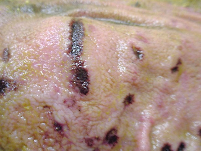
Figure 2. Hemorrhagic abomasal ulcers.
Adapted from Smith (2023), MSD Veterinary Manual.
3.3. Disruption of abomasal emptying
Damage to the gastrin-producing G cells of the pyloric antrum disrupts the hormonal signaling that controls HCl secretion and the rate of chyme passage into the duodenum. The result is irregular abomasal emptying: episodes of excessive retention, alternating with premature passage of insufficiently acidified chyme into the small intestine. This impairs subsequent enzymatic digestion and reduces the absorption of essential amino acids (Fink-Gremmels, 2008; Gallo et al., 2024).
This digestive failure is compounded by the escape of ochratoxin A (OTA). When intake levels or an accelerated passage rate exceed the rumen’s hydrolysis capacity, intact OTA reaches the abomasum and inhibits gastric epithelial renewal. Combined with the low pepsin activity resulting from prior glandular damage, this produces incomplete digestion of microbial protein, drastically reducing the availability of amino acids for production (Fink-Gremmels, 2008).
A critical physiological aspect is the transit of zearalenone (ZEN) and its main ruminal metabolite α-zearalenol (α-ZEL). Upon reaching the abomasum, the acidic pH (2.0-3.5) can alter the solubility of these estrogenic compounds. Since ruminal degradation of ZEN is incomplete and often results in activation rather than detoxification, the abomasum serves as the final conduit for its massive intestinal absorption (Hartinger et al., 2023). The disruption of abomasal emptying induced by other toxins such as DON can prolong the residence time of ZEN in this compartment, increasing the risk of more efficient systemic absorption of its more potent metabolites (Fink-Gremmels, 2008).
3.4. AFM1 transfer to milk: food safety implications
AFB1 is metabolized in the bovine liver into aflatoxin M1 (AFM1), which is actively secreted into milk. Given its mutagenic and carcinogenic potential for humans, its presence in raw and processed milk is subject to strict maximum limits under international legislation: 0.05 µg/kg in the European Union and 0.5 µg/kg in the United States (FDA). This makes the management of mycotoxins in the ruminant’s forestomachs not only a matter of animal welfare and productivity, but also a direct public health issue. The relationship between the concentration of AFB1 in the diet and AFM1 levels in milk is predictable and quantifiable; the transfer rate ranges between 0.3 and 6.2%, although in certain cases it can exceed 7% (Cassel et al., 2001; Kemboi et al., 2020).
4. The abomasum as the final station of gastrointestinal damage from mycotoxins
The abomasal lesion induced by mycotoxins does not occur in isolation: it is the final station of a progressive process of damage that began in the rumen (dysbiosis, reduced VFAs, epithelial permeability), spread to the reticulum (depressed motility and rumination, predisposition to SARA) and to the omasum (laminar necrosis, loss of water and electrolyte absorption), and culminates in the abomasum, compromising the chemical and enzymatic digestion of the ruminant’s true stomach. Integrating these mechanisms into a sequential and cumulative damage model is essential for understanding the pathophysiology of gastrointestinal mycotoxicosis and for designing effective prevention and treatment strategies (Gallo et al., 2024; Antonissen et al., 2014).
Conclusion
The abomasum is the terminal target of mycotoxins in the ruminant’s gastric system. Its cytotoxic effects on glandular secretory cells, ulcerative effects on the mucosa, and disruptive effect on gastrin-HCl signaling compromise protein digestion, impair the absorption of essential amino acids, and, through the transfer of AFM1, put milk safety at risk.
To address this challenge, the use of bioprotective solutions for mycotoxin mitigation is imperative, as they act by preserving glandular function and reducing the bioavailability of toxins such as AFB1. The design of management programs that incorporate this compartmentalized-damage perspective is the most coherent course of action for protecting animal health and food safety.