Introduction
Mycotoxins are secondary metabolites produced by fungi, which contaminate a wide variety of agricultural products, including cereals and forages intended for animal feed. Their presence in the food chain represents a constant threat, as they exert toxic effects and cause substantial economic losses in the agricultural sector.
The toxic effects of mycotoxins are highly diverse and are determined by the type of mycotoxin, the affected species, and other factors (Table 1). Their hepatic and intestinal toxicity has been widely studied, however, their capacity to cause alterations in the development of embryos and fetuses, known as teratogenesis, has received less attention (Chiminelli et al., 2022; Lumsangkul et al., 2019; Silva et al., 2024).
The main pathway by which mycotoxins affect prenatal development is maternal exposure. Mycotoxins such as ochratoxin A (OTA), aflatoxins and deoxynivalenol (DON) have the capacity to cross the placental barrier and reach fetal tissues, compromising fetal development and morphogenesis (Li et al., 2025; Malir et al., 2014; Yu et al., 2017). These compounds exert their teratogenic effects through various toxicity mechanisms, such as the induction of oxidative stress, the activation of apoptosis pathways, or the alteration of DNA, RNA and protein synthesis (Li et al., 2025; Silva et al., 2024).
Aflatoxins
Exposure to aflatoxins, especially to aflatoxin B1 (AFB1), is associated with an increased fetal mortality rate, lower intrauterine growth and the appearance of malformations. The main mechanisms involved in these processes are: increased production of reactive oxygen species (ROS), apoptosis, ferroptosis and the induction of epigenetic modifications that alter fetal gene expression (Li et al., 2025; Popescu et al., 2022; Shin et al., 2018).
In swine, the exposure of early embryos to AFB1 severely compromises their development and implantation. Specifically, it has been reported that exposure to 1 nmol/L AFB1 for 168 h resulted in a reduction in blastocyst formation, resulting in a blastocyst formation rate of 19.01 ± 1.06% compared to 40.13 ± 2.10% in the control group. These findings are thought to be associated with an increase of ROS generation, as this same study reported a relative ROS fluorescence intensity of 1.75 in the AFB1-exposed group, compared to 1.0 in the control group. Thus, oxidative stress would be one of the toxic mechanisms by which AFB1 alters embryonic development.
On the other hand, exposure to this mycotoxin also induces an increase in apoptotic rate (increasing from 5% in the control group, to 15% in the exposed group) (Shin et al., 2018). Furthermore, exposure to 50 µmol/L AFB1 has been linked to lower oocyte maturation rates, as it arrests their maturation at the germinal vesicle breakdown or meiosis I stage, inducing their apoptosis (Popescu et al., 2022).
Among livestock species, poultry exhibit the strongest teratogenic response to aflatoxins. When exposure to AFB1 occurs directly through an in ovo injection into the air cell of fertilized chicken eggs, it triggers drastic toxicity and severe congenital malformations. After 21 days of incubation, doses of 5, 10 or 20 ng per egg cause a drop in the hatching rate (reaching values of 4.76%, 5.48% and 6.9% respectively, compared to 97% in the control), increase embryonic mortality, reduce birth weight (down to 7.40 g compared to 37.30 g in the control) and induce severe morphological abnormalities such as growth retardation, brain deformation, ocular invagination, anophthalmia, beak deformities… (Figure 1) (Ali et al., 2019).
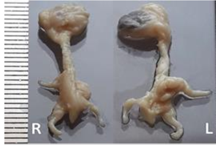
Figure 1. Photograph of a 21-day-old chicken embryo treated with 10 ng of AFB1, with right (R) and left (L) perspectives, showing growth retardation and deformations (Ali et al., 2019).
Conversely, indirect maternal exposure through an in vivo feeding study in laying hens treated with 5 and 10 µg/g for 4 weeks generated a severe clinical impact on reproduction, but without visible physical malformations in the embryos. The damage manifested mainly through an extremely high embryonic mortality that markedly reduced the hatchability of fertile eggs, reaching lows of 21-22% at the highest dose. Additionally, a decrease in the weight of the offspring was recorded by the third week (Howarth and Wyatt, 1976).
At the cellular and molecular level, in vitro models with chicken liver cell lines reveal that AFB1 alters gene expression, resulting in marked reductions in the mRNA of essential target genes (Popescu et al., 2022).
Regarding ruminants, evidence regarding the appearance of visible structural malformations remains scarce at present. In a study with bovine zygotes and embryos, exposure to AFB1 at a concentration of 40 μg/L altered early development, reducing the cell division rate from 67.9% in the control group to 59.4%. After 7.5 days of culture, the same dose reduced the blastocyst formation rate from 19.7% to 8.1% relative to oocytes, and from 30.3% to 14.3% relative to cleaved embryos, reaching total inhibition at doses of 400 and 4000 μg/L. The mechanism behind this alteration of embryonic development is closely linked to oxidative stress, evidenced by a significant increase in ROS production in the exposed zygotes (Jiang et al., 2019).
Fumonisins
Fumonisins, and particularly fumonisin B1 (FB1), exert embryotoxic and teratogenic effects whose scope and severity vary depending on the species and the administered dose. These mycotoxins are capable of causing embryonic death, developmental retardation and severe malformations, such as neural tube defects. These effects are frequently associated with maternal toxicity and are primarily driven by disruption of sphingolipid biosynthesis and interference with folate-related metabolic pathways (Lumsangkul et al., 2019; Silva et al., 2024).
In swine, these effects manifest at the cellular, reproductive and systemic levels. In this regard, it has been confirmed that FB1 has the capacity to cross the placental barrier in mammals, triggering a disruption in both maternal and fetal sphingolipid metabolism. This phenomenon increases the incidence of embryonic resorptions, causing growth retardation and incomplete organogenesis (Silva et al., 2024).
On the other hand, poultry are highly sensitive to the teratogenic effect of fumonisins. FB1 causes growth retardation, incomplete organogenesis and characteristic anatomical malformations such as: hydrocephalus, enlarged beaks, and elongated necks. In ovo assays reflect the extreme sensitivity of fertile eggs as air cell inoculations with purified FB1 doses (0.72 and 72 mg/kg) or combined with fumonisin B2 (FB2), resulted in embryonic mortality rates ranging from 50% to 100%. In fact, microinjections of as little as 0.25 mg/kg of FB1 raised mortality to 56% by day 18, compared to 4% in the control group (Lumsangkul et al., 2019).
Furthermore, surviving embryos show systemic damage to vital organs such as the liver, kidneys and brain. It is known that, after hatching, FB1 perpetuates the productive impact on chickens, ducks, and turkeys, drastically reducing weight gain and causing the atrophy of key immunological organs such as the bursa of Fabricius, thymus, and spleen (Lumsangkul et al., 2019). This immunological atrophy is responsible for rendering animals chronically more susceptible to secondary diseases.
Zearalenone
Zearalenone (ZEN) has potent reproductive toxicity due to its estrogenic activity. This mycotoxin produces structural malformations and direct teratogenicity in some species, whereas in other species these effects are limited to morphological modifications associated with secondary sexual characteristics.
ZEN affects swine with greater severity than other livestock species. It profoundly alters the uterine environment, compromising the viability and quality of the offspring. Exposed sows produce significantly smaller litters (5.7 to 6 piglets in the groups exposed to 50 and 25 mg/kg, respectively, compared to 9 in the control group) and with a lower birth weight (down to 1.16 kg in the group exposed to 25 mg/kg, compared to 1.59 kg in the control).
These toxic effects of ZEN translate directly into an increase in weak-born piglets and a loss of productivity in the farrowing house. Furthermore, this adverse uterine environment has been directly associated with congenital anomalies, documenting skeletal malformations in the carpal and tarsal bones of neonates, accompanied by dermal cysts (Chang et al., 1979).
Ochratoxin A
The primary mechanisms of teratogenic toxicity of OTA are interference with protein and DNA synthesis, the induction of severe oxidative stress, and apoptosis in developing embryonic cells (Pfohl-Leszkowicz and Manderville, 2007; Silva et al., 2024).
In swine, despite being the most sensitive species to OTA, this mycotoxin is not considered a potent teratogen, as it fails to cross the placental barrier efficiently (Pfohl-Leszkowicz and Manderville, 2007).
Conversely, for poultry, OTA is a highly teratogenic and embryotoxic agent. In ovo inoculation assays in fertile eggs demonstrate the impact of the mycotoxin in early stages. Doses starting from 0.05 µg of OTA per egg reduce hatchability to levels between 31% and 38%, compared to 80% in the controls (Zahoor-ul-Hassan et al., 2012). At 53 hours of incubation, exposed embryos already show a critical reduction in the diameter of the optic cup (188.6 µm in the group exposed to 1.00 µg of OTA compared to 329.2 µm in the control) and the lens (81.9 µm in the group exposed to 1.00 µg of OTA compared to 243.9 µm in the control). As incubation progresses, the incidence of malformations increases markedly, with reported cases of anophthalmia, mandibular hypoplasia, microphthalmia, reduced body size, maxillary retrognathism, neural tube closure defects or spina bifida, and exencephaly in the group exposed to 1.00 µg of OTA (Zahoor-ul-Hassan et al., 2012).
The adverse effects of OTA persist after hatching and during the growth phase. Surviving chicks evaluated at day 7 of life exhibit severe pathological alterations: hepatomegaly, renal hypertrophy, and severe atrophy of the bursa of Fabricius, the weight of which drops to less than half (Zahoor-ul-Hassan et al., 2011).
Deoxynivalenol
DON is capable of inducing developmental toxicity during early stages. This negative impact is especially evident in swine, where exposure to 10 µmol/L of DON causes a reduction in normal follicles and an increase in pyknotic oocytes in ovarian tissue, while decreasing viability in cultured swine embryos (Gerez et al., 2017).
Regarding its teratogenic effects in other species, the studies conducted so far remain scarce.
T-2 toxin
Despite the fact that its study is still limited, T-2 toxin has been shown to possesses embryotoxic and developmental toxic potential. In laying hens, the ingestion of T-2 toxin for eight weeks generates a dose-dependent impact on hatchability. The inclusion of levels starting from 2 mg/kg in the diet decreases the hatching rate, which drops to 89.38% upon reaching 8 mg/kg, compared to 96.85% in the control group. Likewise, chicks hatched from breeders exposed to doses of 8 mg/kg show a marked delay in their postnatal development, showing significantly lower body weights at 21 days of age (218.7 g compared to 225.5 g for the controls) (Chi et al., 1977).
| MYCOTOXIN | ANIMAL SPECIES | DOSE/EXPOSURE | MAIN EFFECTS | REFERENCES |
| Aflatoxin B1 | Swine (early embryos) | 1 nmol/L 168 h In vitro | Reduction in blastocyst formation and cellular apoptosis. | Shin et al. (2018) |
| Aflatoxin B1 | Swine (oocytes) | 50 µmol/L In vitro | Arrest of oocyte maturation. | Popescu et al. (2022) |
| Aflatoxin B1 | Poultry (chicken eggs) | 5, 10 or 20 ng/egg 21 days of incubation In ovo | Reduction in hatching rate, increase in mortality, reduction in weight, induction of morphological abnormalities. | Ali et al. (2019) |
| Aflatoxin B1 | Poultry (laying hens) | 5 or 10 µg/g 4 weeks In vivo | High embryonic mortality, reduction in hatchability of fertile eggs and offspring weight. | Howarth and Wyatt (1976) |
| Aflatoxin B1 | Bovine (zygotes and embryos) | 40 µg/L 7.5 days In vitro | Reduction in cell division rate and blastocyst formation rate. | Jiang et al. (2019) |
| Fumonisin B1 | Poultry (chicken eggs) | 0.25 – 72 mg/kg 21 days of incubation In ovo | Increase in embryonic mortality, damage to liver, kidneys, and brain in embryos, reduction in weight gain, and atrophy of immunological organs in animals after hatching. | Lumsangkul et al. (2019) |
| Zearalenone | Swine (multiparous sows) | 25, 50 or 100 mg/kg 183 days In vivo | Reduced litters with lower birth weight and congenital anomalies. | Chang et al. (1979) |
| Ochratoxin A | Poultry (chicken eggs) | 0.05 – 1.00 µg/egg 22 days of incubation In ovo | Decrease in hatchability, alterations in the optic cup and lens, increased incidence of malformations, hepatomegaly, renal hypertrophy, and atrophy of the bursa of Fabricius. | Zahoor-ul-Hassan et al. (2012) |
| Deoxynivalenol | Swine (ovaries) | 10 µmol/L 48 h Ex vivo | Reduction in the number of normal follicles and increase in pyknotic oocytes at all stages of follicular development. | Gerez et al. (2017) |
| T-2 toxin | Poultry (laying hens) | 2, 4 or 8 mg/kg 8 weeks In vivo | Reduction in hatching rate and delay in postnatal development of chicks. | Chi et al. (1977) |
Table 1. Summary of the reviewed literature.
Conclusions
Mycotoxin-induced teratogenesis represents a serious risk to animal health, welfare, and livestock sector productivity. However, the available scientific evidence highlights the need for more information and a greater research effort are needed in this area.
Currently, there are critical gaps in scientific knowledge, with limited information available regarding the in vivo teratogenic effects in ruminant livestock, as well as the true pathological impact of mycotoxins such as deoxynivalenol and T-2 toxin during the gestational period.